


 Bandra
 Kandivali
 Juhu
Juhu
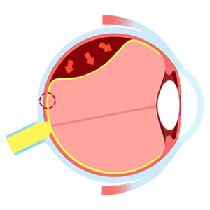
What Is Retinal Detachment?
Retinal detachment is a serious eye problem that usually does not cause pain. It occurs when the retina (the light-sensitive layer at the back of the eye) separates from its normal position. This can affect your vision and may lead to permanent vision loss if not treated quickly.
The retina helps you see by sensing light and sending signals to the brain. When it detaches, it cannot function properly, leading to blurred or lost vision.
Seek immediate medical help if you notice:
- A sudden increase in floaters
- Flashes of light
- A shadow or curtain over your vision
These may be warning signs of retinal detachment. Do not wait for pain - this is a medical emergency and early treatment is essential to protect your vision. The condition often begins with mild symptoms like floaters or flashes but can progress quickly and lead to permanent vision loss if not treated on time.
What Are the Retinal Detachment Symptoms You Should Not Ignore?
Retinal detachment is a serious eye emergency that can lead to permanent vision loss if not treated immediately. Symptoms may appear suddenly or develop slowly, depending on how much of the retina is affected. It is important to recognize the warning signs early.
Common signs include:
- Sudden increase in eye floaters: Floaters are small dots, specks or thread-like shapes that move in your vision. While common with age, a sudden increase in floaters can indicate a problem and should not be ignored.
- Colors appear faded or dull: If things around you look less bright or colors seem faded, it may be a sign of retinal problems. The retina helps you see colors clearly, so any change in how colors appear should not be ignored. It is important to get your eyes checked if you notice these symptoms.
- Blurred vision: Vision becomes unclear or distorted. Objects may appear wavy, bent or blurred. Straight lines may not look straight, making it hard to read or focus on details.
- Persistent double vision: Seeing double for a long time should not be ignored. While temporary double vision can happen due to tiredness, ongoing double vision may be linked to problems with the retina or other vision-related conditions.
- Flashes of light: You may notice brief flashes of light, especially in your side vision. These can look like quick flickers or lightning-like streaks. If they happen often or increase, it may be a warning sign.
- Shadow or curtain: You may feel like a dark shadow or curtain is moving across your vision, often starting from the side and spreading inward. This is a serious sign and requires immediate care.
- Ring of floaters: Sometimes, floaters may appear in a ring-like pattern near your central vision. This can be an early sign of a retinal tear and needs quick attention.
- Central vision loss: In advanced stages, central vision may become blurred or lost, especially if the main part of the retina (macula) is affected. This can lead to significant vision problems if not treated quickly.
- Veil-like effect: Some people experience a blurry or covered feeling, as if a veil is over their eyes. This may worsen over time if not treated.
- Loss of side vision: Loss of side (peripheral) vision, also called “tunnel vision,” can be a sign of serious retinal damage. If you start bumping into things or find it hard to see from the corners of your eyes, you should visit an eye specialist.
If you notice any of these symptoms, seek medical help immediately. Early diagnosis and treatment can help protect your vision.
When Should You See an Eye Specialist for Retinal Detachment?
You should visit an eye specialist within 24 hours if you notice sudden changes in your vision. Retinal detachment is a medical emergency and early treatment is very important to prevent permanent vision loss.
Do not delay if you notice any sudden changes in vision, like the following:
- Flashes of light: Seeing sudden flashes like lightning or sparks
- New or increased floaters: A sudden rise in dots, specks or cobweb-like shapes
- Shadow or curtain effect: A dark shadow or curtain covering part of your vision
- Sudden vision changes: Quick blurring or loss of vision in one eye
If you experience any of these symptoms, do not ignore them and seek medical help immediately.
What Causes Retinal Detachment?
Retinal detachment occurs when the retina separates from its normal position at the back of the eye. This can occur due to aging changes in the eye, injury or severe nearsightedness. It happens in three main types - by tears or holes, pulling by scar tissue or fluid buildup. And it often needs urgent surgery to prevent permanent vision loss.
Common causes of retinal detachment include:
- Aging (Posterior Vitreous Detachment - PVD): With age, the gel inside the eye shrinks and can pull on the retina, sometimes causing a tear.
- Eye injury: Any strong hit or trauma to the eye can cause the retina to tear or detach. Wearing protective glasses during sports or risky activities can help prevent this.
- Severe myopia (nearsightedness): In high myopia, the eyeball is longer, which stretches and thins the retina. This makes it easier for tears to develop. Regular eye checkups are important to monitor retinal health.
- Previous eye surgery: After surgeries like cataract or glaucoma, some people may have a higher risk of retinal detachment. Changes inside the eye can sometimes lead to retinal tears or fluid buildup. If you notice flashes or floaters after surgery, seek medical help quickly.
- Lattice retinal degeneration: This condition makes the outer retina thin and weak, increasing the chance of tears and detachment. Regular eye exams can help detect it early.
- Diabetes (diabetic retinopathy): In people with diabetes, damaged blood vessels and scar tissue can pull the retina away from its position. Good sugar control and regular eye checkups can help reduce the risk.
- Family history: If retinal detachment runs in your family, your risk may be higher. Regular eye screening is recommended in such cases.
In many cases, retinal detachment can develop due to more than one cause. A mix of these factors may increase the risk, so regular eye check-ups are important for early detection and timely treatment.
What Are the Risk Factors for Retinal Detachment?
Certain factors can increase the chances of retinal detachment. Some people may have a higher risk due to family history, while others may develop it due to injury or eye conditions.
Common risk factors include:
- Aging: As you age, the gel inside the eye shrinks and may pull on the retina, causing tears. The risk increases after 50.
- High myopia (nearsightedness): A longer eye shape makes the retina thin and more likely to tear.
- Retinal detachment in one eye: If you have had detachment in one eye, the risk is higher in the other eye. Regular checkups are important.
- Previous eye surgery: Surgeries like cataract removal can increase the risk, as they may cause changes inside the eye.
- Eye injury: A strong hit or injury to the eye can cause the retina to tear or detach.
- Other eye diseases: Conditions like uveitis or lattice degeneration can weaken the retina and increase the risk.
- Family history: If someone in your family has had retinal detachment, your risk may be higher.
Regular eye check-ups are important if you fall into these categories, as early diagnosis and timely treatment can help prevent permanent vision loss.
Did You Know?
- Retinal detachment is painless but can cause sudden vision loss.
- It usually affects one eye but can occur in both.
- Floaters and flashes are often the first warning signs.
- It is a treatable condition if diagnosed early.
- Delay in treatment can lead to permanent blindness.
What Are the Different Types of Retinal Detachment?
Retinal detachment is a serious condition where the retina separates from the tissue beneath it. It is mainly divided into three types based on the cause and all types need immediate treatment to prevent permanent vision loss.
The three main types of retinal detachment, depending on the cause:
Rhegmatogenous Retinal Detachment
This is the most common type. It happens when the gel inside the eye pulls on the retina and creates a tear or hole, allowing fluid to pass underneath.
Risk factors include:
- Aging
- Eye injury
- Family history
- High myopia (nearsightedness)
- Retinal tear or detachment in the other eye
- Previous cataract surgery
Tractional Retinal Detachment
This type occurs when scar tissue forms on the retina and pulls it away from the back of the eye. It is usually linked to abnormal blood vessel growth.
Common causes include:
- Diabetic retinopathy
- Sickle cell disease affecting the eye
- Scar tissue formation inside the eye
- Eye infections or inflammation
- Retinopathy of prematurity (ROP)
Exudative (Serous) Retinal Detachment
This type happens when fluid collects under the retina without any tear or hole. The fluid buildup pushes the retina away from its normal position.
Possible causes include:
- Age-related macular degeneration
- High blood pressure
- Severe eye inflammation
- Pregnancy-related conditions (like preeclampsia)
- Coats disease
- Eye tumors
All types require timely diagnosis, quick medical care and appropriate treatment to restore and protect vision.
How Is Retinal Detachment Diagnosed?
Retinal detachment is diagnosed through a detailed eye examination by an eye specialist. The doctor usually performs a dilated eye exam to clearly see the retina and check for any tears or detachment. Early diagnosis is very important to prevent permanent vision loss.
Common diagnostic tests include:
- Dilated eye exam: Eye drops are used to widen the pupil so the doctor can examine the back of the eye with a bright light and specialized lenses.
- Slit-lamp exam: It is used to check the vitreous for tiny pigment particles, called “tobacco dust” or Shafer’s sign, which may indicate a retinal tear.
- Eye ultrasound (B-scan): It is used when bleeding or cataracts block a clear view of the retina, helping to create images of the inside of the eye.
- Indirect ophthalmoscopy: In this test, a special light and lens are used to check for retinal tears or detachment. Sometimes, gentle pressure is applied on the eyelid to see the outer edges of the retina more clearly.
- OCT (Optical Coherence Tomography): It is a painless scan that provides detailed images of the retina and helps detect fluid buildup under it.
- Fundus imaging: During this test, wide-angle photos of the retina are taken to get a clearer and detailed view. Usually your eyes are dilated for this test.
- CT scan (Computed Tomography): This scan may be done in case of any history of trauma or possible penetrating eye injury to check internal damage.
These tests are safe and help doctors diagnose the condition accurately and start treatment quickly.
What Are the Complications of Retinal Detachment?
Retinal detachment is a serious condition that can cause permanent vision loss if not treated on time. In some cases, even after treatment, complications may occur. Early care is important to protect your vision.
Possible complications:
- Permanent vision loss: It occurs especially if the central part of the retina (macula) is affected.
- Scar tissue (PVR): Scar tissue may form on the retina and cause it to detach again.
- Glaucoma: It happens with increased pressure inside the eye due to long-term damage or after surgery.
- Cataract: Clouding of the eye lens may happen, which may develop after treatment.
- Vitreous hemorrhage: Bleeding inside the eye can even happen.
- Epiretinal membrane: In this a thin layer forms on the retina, leading to distorted vision.
- Low eye pressure (hypotony): Sometimes abnormally low pressure is noted inside the eye.
- Infection: It is a rare but serious complication after surgery.
Timely diagnosis and treatment can help reduce the risk of these complications and preserve vision.
What Are the Available Retinal Detachment Treatment Options?
Retinal detachment is a medical emergency and needs quick treatment to save vision. Treatment depends on the severity and type of detachment. Early cases may be managed without major surgery.
Surgery is usually required to reattach the retina and prevent permanent vision loss. In many cases, these treatments are highly successful.
Common treatment options include:
Vitrectomy
The most commonly used surgery, especially done for complex cases, where the gel inside the eye is removed and replaced with a gas bubble or silicone oil to hold the retina in its proper position.
- Removes the gel pulling the retina
- Replaces it with gas or oil
Pneumatic Retinopexy
A gas bubble is injected into the eye to push the retina back, often used in simpler cases and may require specific head positioning for several days.
- Gas bubble injected into the eye
- Pushes retina back into place
Scleral Buckling
A small band is placed around the eye to gently push the retina back into its normal position.
- A band supports the eye wall
- Helps reattach the retina
Laser Therapy or Cryotherapy
These methods use heat (laser) or freezing to seal retinal tears and help attach the retina properly.
- Used for early retinal tears
- Helps seal the retina
Early diagnosis and timely treatment give the best chance of restoring and protecting your vision. Your eye specialist will choose the most suitable treatment based on your condition to ensure the best possible result.
What Is Retinal Detachment Surgery, How Does It Work and What Are Its Benefits?
Retinal detachment surgery is an emergency procedure done to reattach the retina and save vision. It has a high success rate and early treatment can help prevent permanent vision loss.
How the Surgery Works
Doctors use one or a combination of different methods to fix the retina and keep it in place. These procedures are usually done under local or general anesthesia.
How it works:
- The retina is repositioned to its normal place.
- Tears are sealed using laser or freezing methods.
- Support is provided using gas, oil or scleral buckle.
These treatments help reattach the retina and restore stable vision when done on time.
Key benefits:
- Prevents permanent vision loss
- Restores or improves vision
- Stops further retinal damage
- High success rate with timely treatment
Most procedures are done under local anesthesia and may be day-care or short-stay procedures.
What Are the Different Types of Retinal Detachment Surgery Available?
Different surgical methods are used depending on the condition:
Pneumatic Retinopexy
- Gas bubble injected into the eye
- Helps push retina back into place
- Suitable for simple detachments
Scleral Buckling
- Silicone band placed around the eye
- Relieves pressure on the retina
- Effective for many types of detachments
Vitrectomy
- Removes vitreous gel pulling the retina
- Retina is reattached and supported
- Used for complex or severe cases
Your eye doctor will decide the most suitable procedure for you.
When Can You Resume Activities After Retinal Detachment Treatment?
Following your doctor’s advice carefully helps ensure a smooth recovery and better vision results.
- Office work: Light office work can usually be resumed in 1 to 2 weeks, while heavy work may take 4 weeks or more.
- Light exercise: Simple activities like walking are generally safe and encouraged.
- Heavy activities: Avoid heavy lifting, gym workouts, swimming or sports for at least 4 to 6 weeks.
- Flying: Not allowed for some time if a gas bubble is used, as it can expand at high altitudes.
- Driving: You can start driving after 1 to 2 weeks, only if your doctor confirms your vision is safe.
Taking these precautions helps ensure proper healing and better vision recovery.
Key Points to Know:
- Retinal detachment is a medical emergency.
- Early treatment gives better results.
- Surgery success depends on timing.
- Vision recovery may take time.
- Regular follow-up is important.
When Should I Get Retinal Detachment Treatment?
Retinal detachment is a medical emergency and needs immediate treatment, ideally within 24 to 48 hours - to prevent permanent vision loss. If you notice any sudden changes in your vision, see an eye specialist right away.
Warning signs that need urgent care:
- Sudden flashes of light
- A sudden increase in floaters or black spots
- A dark shadow or curtain over your vision
- Sudden blurred or reduced vision
It is always best to consult your eye specialist without delay and follow regular check-ups. Do not wait too long, as quick action can help save your vision and prevent serious complications.
How to Prepare for Retinal Detachment Surgery?
Preparing well for surgery can help ensure a smooth procedure and faster recovery. Since this surgery is important to protect your vision, following the doctor’s instructions carefully is essential.
- If you have diabetes: Do not take insulin on the morning of surgery unless your doctor advises it. Carry your medicines with you.
- If you feel unwell: Inform your doctor immediately if you have fever, cold or any illness before surgery, as it may need to be postponed.
- Mental preparation: Learn about the procedure beforehand to feel more comfortable and reduce anxiety.
How to Prepare Before Retinal Detachment Surgery?
- Inform your doctor: Tell your doctor about all medicines you are taking, especially blood thinners, as some may need to be stopped before surgery.
- Fasting: Do not eat or drink anything after midnight before the surgery.
- Arrange transport: Plan for someone to take you home, as you will not be able to drive after the procedure.
- Hygiene and clothing: Take a bath and wash your hair the night before. Wear loose, comfortable clothes and avoid makeup, lotion or jewelry.
- Prepare your home: Set up a comfortable space for recovery and keep easy meals ready.
- Head positioning support: If a gas bubble is used, you may need to keep your head in a certain position. Arrange pillows or special support in advance.
Proper preparation can help reduce stress and support a better recovery after surgery.
What Happens During Retinal Detachment Surgery?
- Procedure is done under local or general anesthesia.
- Surgery duration varies (30 minutes to a few hours).
- You may go home the same day or after a short
- Vision may be blurred initially.
What to avoid before surgery?
- Do not wear contact lenses on the day of surgery.
- Avoid smoking and alcohol for at least 24 hours before the procedure.
- Do not plan air travel or high-altitude travel if a gas bubble may be used.
How to Care After Retinal Detachment Surgery?
Proper care after surgery is very important for healing and protecting your vision. You may need to follow specific instructions, especially if a gas bubble is used in your eye.
- Head positioning: You may need to keep your head in a fixed position (often face-down or sideways) for 1 to 2 weeks to help your retina heal properly.
- Eye protection: Wear an eye shield while sleeping to avoid rubbing the eye. Keep water, soap or dust away from the eye.
- Activity restrictions: Avoid heavy lifting, bending, exercise and sudden movements for a few weeks to prevent pressure on the eye.
- Use of medicines: Use prescribed eye drops regularly to prevent infection and reduce swelling.
- Driving and travel: Do not drive until your doctor allows it. Avoid air travel if a gas bubble is present in the eye.
Following these instructions carefully helps ensure better healing and recovery.
Will My Vision Fully Recover After Retinal Detachment?
Vision recovery depends on how early the treatment is done.
- Early treatment gives better outcomes
- Some vision loss may be permanent in severe cases
- Recovery may take weeks to months
- Regular follow-up helps monitor healing
What Are the Possible Risks and Complications of Retinal Detachment Treatment?
Though treatment is effective, some risks may include:
- Infection
- Bleeding
- Increased eye pressure
- Cataract formation
- Re-detachment of retina
Timely care and follow-up reduce these risks.
Contact Your Doctor Immediately If:
- Severe or increasing eye pain
- Sudden drop in vision
- New flashes of light or more floaters
- Unusual discharge or swelling
What Happens If Retinal Detachment Is Not Treated?
If retinal detachment is left untreated, it can lead to serious complications, like:
If retinal detachment is not treated on time, it can lead to permanent and severe vision loss or even complete blindness in the affected eye. This happens because the retina stops getting proper blood supply and its cells start to die within hours or days.
- Permanent vision loss: The retina cannot work without blood supply, leading to irreversible loss of vision.
- Dark shadow in vision: A shadow or “curtain” may start from the side and slowly cover the entire field of vision.
- Loss of central vision: If the central part of the retina (macula) is affected, sharp vision is lost and may not return.
- Scar tissue formation (PVR): Scar tissue can develop, making the retina stiff and harder to treat.
- Other eye problems: Conditions like high eye pressure (glaucoma) or cataracts may develop
- Shrinkage of the eye: In advanced cases, the eye may gradually shrink and lose function.
Note: Early treatment is important. Acting quickly can help save your vision and prevent these serious complications.
How Long Does It Take to Recover After Retinal Detachment Treatment?
Recovery after retinal detachment treatment usually takes a few months. Most people can return to daily activities within 2 to 4 weeks, but full healing may take 3 to 6 months or longer. Vision is often blurry at first and improves gradually over time.
- First 1 to 2 weeks: Avoid bending, heavy lifting or exercise. You may need to wear an eye shield, especially while sleeping.
- Head positioning: If a gas bubble is used, you may need to keep your head in a specific position (face-down or side) for about 1 to 2 weeks to help the retina heal properly.
- Vision improvement: Vision is usually poor just after surgery but slowly gets better over a few months. In some cases, it may take up to a year.
- Follow-up visits: Regular check-ups are important to make sure the retina stays in place and heals well.
- Warning signs: Contact your doctor immediately if you have severe pain, new flashes of light or sudden worsening of vision.
Regular follow-ups are important for proper healing.
What Questions Should You Ask Your Eye Specialist?
You can ask your doctor these simple questions:
- Do I have retinal detachment?
- How serious is my condition?
- Which type do I have?
- What treatment do I need?
- Is surgery required in my case?
- What are the risks and recovery time?
- Can my vision be fully restored?
- How soon should I get treatment?
- What precautions should I follow?
- Will this happen again?
Can I Prevent Retinal Detachment?
You may not always be able to prevent retinal detachment, especially with age, but you can lower your risk and protect your vision by taking the right steps.
Key ways to prevent retinal detachment:
- Regular eye check-ups: Get routine dilated eye exams to detect early problems, especially if you are nearsighted or have a family history.
- Protect your eyes: Wear safety glasses during sports or activities that may cause eye injury.
- Manage health conditions: Control conditions like diabetes to reduce the risk of eye damage.
- Know the warning signs: Do not ignore symptoms like sudden flashes, new floaters or a shadow/curtain in your vision.
If you notice any of these symptoms, seek immediate medical care. Early treatment can help prevent permanent vision loss.
Book Your Retinal Detachment Consultation at Ojas Maxivision Eye Hospital
If you are noticing sudden vision changes, do not ignore them. At Ojas Maxivision Eye Hospital, our experienced eye specialists provide advanced care for retinal conditions with a patient-first approach.
If you are searching for an eye hospital in Mumbai or need timely retinal detachment treatment in Mumbai, our team is here to guide you at every step. Early care can make all the difference - act now to protect your vision.
Frequently Asked Questions About Retinal Detachment
Retinal detachment is usually not painful, which is why many people ignore early symptoms. However, it causes sudden vision changes like flashes of light, floaters or blurred vision. Even without pain, it is a serious eye condition and needs immediate medical attention to prevent vision loss.
The early signs of retinal detachment include a sudden increase in floaters, flashes of light, blurred vision or a shadow or curtain over your vision. These symptoms may appear quickly and should never be ignored, as early treatment can help save your eyesight.
No, retinal detachment cannot heal on its own. It is a serious condition that requires urgent medical or surgical treatment. Delaying care can lead to permanent vision loss, so it is important to consult an eye specialist as soon as symptoms appear.
Yes, retinal detachment is a medical emergency. Immediate treatment is needed to reattach the retina and prevent permanent damage. The sooner you receive care, the better the chances of saving your vision and avoiding complications.
Yes, retinal detachment can affect both eyes. If you have had detachment in one eye, the risk in the other eye may increase, so regular eye check-ups are very important.
Yes, diabetes can increase the risk of retinal detachment, especially if diabetic retinopathy is not well controlled. High blood sugar levels damage the retina over time, which can lead to serious complications, including detachment.
Surgery is not always required in very early cases. Small retinal tears can sometimes be treated with laser or freezing methods. However, most retinal detachments need surgery to properly reattach the retina and restore vision.
Retinal detachment surgery has a high success rate, especially when treated early. The final vision result depends on how quickly treatment is done and whether the central retina was affected.
Yes, retinal detachment can happen again even after successful treatment. However, regular follow-ups and early detection can help manage the condition and reduce the risk of further complications.
The duration of retinal detachment surgery depends on the type and severity of the condition. It usually takes between 30 minutes to a few hours. Your doctor will explain the expected time based on your specific case.
In many cases, retinal detachment surgery is done as a day-care procedure, meaning you can go home the same day. However, some patients may require a short hospital stay depending on their condition and recovery needs.
You should avoid driving after surgery until your doctor confirms that your vision is safe. Vision may be blurry during the initial recovery period, especially if a gas bubble is used in the eye.
You may need glasses after retinal detachment surgery, depending on how your retina heals. Your doctor will check your vision during follow-ups and recommend glasses if needed to improve clarity.
Yes, certain precautions are necessary after surgery. You should avoid heavy lifting, bending, and strenuous activities. If a gas bubble is used, flying or traveling to high altitudes must be avoided until it fully dissolves.
Vision recovery depends on how early the treatment is done. If treated quickly, vision can improve significantly. However, in advanced cases, some vision loss may be permanent, especially if the central retina is affected.
Yes, retinal detachment can occur in children, but it is rare. It is usually linked to eye injuries, genetic conditions or other eye problems. Early diagnosis and treatment are important to protect vision.
People with high myopia (powerful glasses), diabetes, previous eye surgery or eye injury are at higher risk. Family history and aging can also increase the chances, so regular eye check-ups are recommended.
No, glasses cannot treat retinal detachment. It is a medical condition that requires proper treatment such as laser therapy or surgery. Glasses may help vision after treatment but cannot fix the detachment itself.
Yes, if not treated on time, retinal detachment can lead to permanent vision loss or blindness. Early diagnosis and immediate treatment are essential to save eyesight and prevent serious complications.
The cost of retinal detachment treatment varies depending on the type of surgery, severity of the condition and hospital facilities. It is best to consult the hospital directly for an accurate estimate based on your case.
Yes, Ojas Maxivision Eye Hospital provides cashless treatment with selected insurance companies. It is empaneled with CGHS, RBI, NABARD and BSNL. Please contact the hospital to check your eligibility and complete the required documents before particular eye treatments.
Ojas Maxivision Eye Hospital has multiple centers in Mumbai, Maharashtra, including Bandra (West), Kandivali (East) and Juhu. You can choose the nearest location and book an appointment for consultation and treatment.
Key Facts About Retinal Detachment
- It can occur when the retina separates from the back of the eye.
- It is a medical emergency.
- It can cause sudden vision loss if untreated.
- Common signs include flashes, floaters and shadows.
- Causes include aging, eye injury or diabetic retinopathy.
- It is painless but serious.
- Early treatment can save vision.
- Surgery is often required in advanced cases.